| dc.contributor.author | Bolat Gümüş, Esra | |
| dc.contributor.author | Şatır, Samed | |
| dc.contributor.author | Kuştarcı, Alper | |
| dc.date.accessioned | 2023-10-02T06:33:55Z | |
| dc.date.available | 2023-10-02T06:33:55Z | |
| dc.date.issued | 2022 | en_US |
| dc.identifier.uri | https://www.scopus.com/record/display.uri?eid=2-s2.0-85129345090&origin=resultslist&sort=plf-f&src=s&nlo=&nlr=&nls=&sid=20c4fe371b9b908d4f7e419f791331ff&sot=aff&sdt=cl&cluster=scofreetoread%2c%22all%22%2ct&sl=72&s=AF-ID%28%22Alanya+Alaaddin+Keykubat+University%22+60198720%29+AND+SUBJAREA%28MEDI%29&relpos=55&citeCnt=0&searchTerm= | |
| dc.identifier.uri | https://hdl.handle.net/20.500.12868/2363 | |
| dc.identifier.uri | https://academic.oup.com/dmfr/article/51/4/20210512/7261280 | |
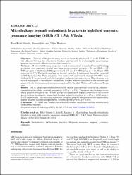
| dc.description.abstract | Objectives: The aim of the present study was to evaluate the effects of 1.5 T and 3 T MRI on the adhesion between the orthodontic brackets and the teeth by evaluating the microleakage between the enamel, adhesive and brackets interfaces. Methods: 58 extracted human premolars which were received a standard bracket bonding procedure were randomly divided into three groups; control group (n = 20; no MRI), 1.5 T MRI group (n = 19; 20 min MRI exposure of 1.5 T) and 3 T MRI group (n = 19; 20 min MRI exposure of 3 T). The teeth were kept in distiled water for 2 weeks, and thereafter subjected to 500 thermal cycles. Then, specimens were sealed with nail varnish, stained with 0.5% basic fuchsin for 24 h, sectioned and photographed under a stereomicroscope. Microleakage was scored with regard to the adhesive–enamel and bracket–adhesive interfaces at the occlusal and gingival levels. Statistical analysis was accomplished by Kruskal–Wallis and Bonferroni–Dunn tests. Results: All of the groups exhibited statistically similar microleakage scores in the adhesive–enamel interface along occlusal margins (p>0.05, p = 0.331). The mean microleakage scores along gingival margins in the 3 T MRI group was significantly higher compared to the control group both in the adhesive–enamel and bracket–adhesive interfaces (p<0.05, p = 0.019 and p = 0.020 respectively). The microleakage scores along the gingival margins were also significantly higher than the occlusal margins in the 3 T MRI group (p[removed] | en_US |
| dc.language.iso | eng | en_US |
| dc.relation.isversionof | 10.1259/dmfr.20210512 | en_US |
| dc.rights | info:eu-repo/semantics/openAccess | en_US |
| dc.subject | Orthodontics | en_US |
| dc.subject | Orthodontic brackets | en_US |
| dc.subject | Magnetic resonance imaging | en_US |
| dc.subject | Artefacts | en_US |
| dc.subject | Adhesive | en_US |
| dc.title | Microleakage beneath orthodontic brackets in high field magnetic resonance imaging (MRI) AT 1.5 & 3 Tesla | en_US |
| dc.type | article | en_US |
| dc.contributor.department | ALKÜ, Fakülteler, Diş Hekimliği Fakültesi, Ağız, Diş ve Çene Radyolojisi Ana Bilim Dalı | en_US |
| dc.identifier.volume | 51 | en_US |
| dc.identifier.issue | 4 | en_US |
| dc.relation.journal | Dentomaxillofacial Radiology | en_US |
| dc.relation.publicationcategory | Makale - Uluslararası Hakemli Dergi - Kurum Öğretim Elemanı | en_US |


